If a risk of chromosome disease in fetus is hgher than populational (women aged above 35, child birth with a chromosome disease or presence of biochemical markers of chromosome disease– typical changes of the levels alpha – fetoprotein and chorionic gonadotropin), prenatal investigation of the fetal karyotype (chromosomal analysis) is recommended. For chromosamal analysis of the fetus fetal cells are required.
Invasive prenatal testing is used to identify fetus chromosome abnormalities (Down’s syndrome, Edwards’ syndrome, etc). Thus karyotyping of cells of chorion/placenta/amniocytes or blood lymphocytes sampling is provided.
Karyotype analysis is a standard method available in our clinic for identifying numerical and structural chromosomal aberrations.
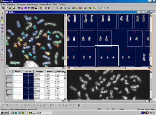
Fig.1 Computer-aided analysis of the karyotype of a fetus with a Down syndrome, performed in our clinic
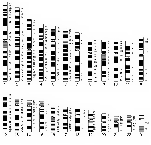
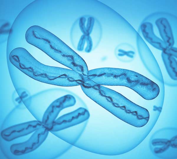
Fig.2 Schematic of chromosome structure using the G-staining method in compliance with the international classification
The chromosome, chorion and placenta fibre cells analyses contribute significantly to the identification of chromosomal anomalies, such aberrations are revealed in 98.6% which helps to take the timely measures.
The up-to-date techniques allowed finding out that many severe congenital abnormalities are caused by numerical and structural chromosomal aberrations. Down syndrome is one of the most common and well-known chromosome abnormalities, being a severe congenital pathology with mental retardation, immunity disorders and heart activity disorders, etc. It occurs because of the presence of an extra 21st chromosome in the karyotype.
Invasive prenatal testing for fetus chromosome abnormalities is recommended in the following cases:
- For pregnant woman aged above 35 years old (e.g. Down’s syndrome generally occurs for 1 in 700 of newborns, but for 1 in 30 of children born by women aged above 35, this is true for other chromosome abnormalities as well);
- Down’s syndrome, other chromosome diseases, or multiple congenital abnormalities in history of a previous child (children);
- Ultrasonic markers of fetus chromosome abnormalities;
- Balanced chromosomal rearrangement in either parent;
- Realtive indication is a result of biochemical screening (alpha – fetoprotein and chorionic gonadotropin) indicating a high risk of Down syndrome.
Invasive methods alow to get a sample of fetal cells, or cells from provisional organs (chorionic villus sampling, placenta) for chromosomal analysis. This procedures is performed through transabdominal biopsy under ultrasound control. Techniques differ depending on pregnancy term.
Invasive methods performed in our clinic include:
Chorion biopsy is a procedure for taking a small piece of placental tissue (chorionic villi) (in 10-14 weeks);
Placenta biopsy is a procedure for taking a small piece of placenta cells (in 14-20 weeks);
Amniocentesis is a puncture of the fetal bladder for taking some amniotic fluid (in 15-18 weeks);
Cordocentesis is a procedure for obtaining blood cells from the umbilical cord (umbilical blood sampling) (after 20 weeks);
In rare cases a fetal tissues biopsy can be performed.
The risk of pregnancy complications (spontaneous termination of a pregnancy or a fetal death) after chorion biopsy and placenta biopsy is 1% which is typical rate for the first trimester; for amniocentesis this rate is even lower - about 0.2%; for cerdocentesis it increases up to 3.3%.
Invasive prenatal techniques are available for the majority of monogenic diseases. All invasive testing techniques are used for this purpose. However, unlike the cytogenetic prenatal diagnostics including the karyotype study, the gene sequences or areas are examined.
In case of monogenic diseases we provide all the invasive techniques for assesment of fetal cells. The samples are sent to professional laboratories specializing in particular molecular diagnostic techniques.
Nowadays in Russia the following testing for monogenic disorders is available:
- Adrenogenital syndrome
- Albinism type OCA 1
- Friedreich\'s ataxia
- Achondroplasia
- Wilson’s disease
- Willebrand’s disease
- Lesch-Nyhan syndrome
- Norry’s disease
- Unterricht-Lunborg’s disease
- Hunter’s disease
- Congenital arachnodactylia
- Congenital myodystrophy, type Fukuyama
- b- thalassemia
- Haemophilia A and B
- Hypophyseal nanosomia
- deficiency of acyl-CoA dehydrogenase of fatty acids with a medium chain length
- deficiency of alfa-1-antitrypsin
- Dushenn-Bekker myodystrophy
- Emery-Dreiyfus myodystrophy
- myotonic myodystrophy
- Tompson-Bekker myotonia
- Cystic fibrosis
- Sharko-Mary-Tut 1st type atrophy
- sensorineural hearing loss
- oculopharyngeal myodystrophy
- pseudoachondroplastic dysplasia
- recessive polycystic renal disease
- familial hypercholesteremia
- Alport\'s syndrome
- Acrosphenosyndactylia
- Coffin-Lowry’s syndrome
- Louis-Bar syndrome
- Martin-Bel’s syndrome
- Marfan’s syndrome
- Miller-Dicker syndrome
- Nijmegen syndrome
- Smitt-Lemly-Opitz syndrome
- Feminizing testes syndrome
- Holt-Omar’s syndrome
- Ehlers-Danlos syndrome (classical type)
- Spastic paraplegia
- Spinal and bulbar Kennedy’s amyotrophy
- Spinal Hoffmann’s, Kugelberg-Welander’s amyotrophy
- Felling’s disease
- Huntington\'s chorea
- Anhydrotic ectodermal dysplasia
- X-linked agammaglobulinemia
- X-linked lymphoproliferative disorder (Duncan’s disease)
Invasive prenatal diagnostics is applied for sex determination and determination of the paternity during the early pregnancy.


